Showing posts with label treatments. Show all posts

Tuesday, April 28, 2015

Guts, Bugs and "Slow Sepsis" in ME/CFS

Last fall, Invest in ME announced an update in its research into the microbiome of ME/CFS patients. The study is based on the hypothesis that exposure to gut microbes through increased intestinal permeability (aka "leaky gut") initiates microbe-driven inflammatory reactions in ME/CFS. The researchers hope to detect increases in serum antibodies towards specific intestinal bacteria.

The goal of the study is "to determine if alterations in intestinal barrier function and/or microbiota firstly, exists in ME/CFS patients and secondly, whether there is an interaction between microbe-driven inflammatory responses and neuronal proteins."

As a hypothesis, this study has a firm foundation. 

Antibodies produced by the infiltration of gut bacteria into the bloodstream can initiate, among other things, a cascade of potentially lethal inflammatory responses. In 2014, researchers at the University of Chicago found that patients in the Intensive Care Unit had only limited kinds of gut flora left in their intestines after multiple rounds of antibiotics. Not surprisingly, these were flora that were pathogenic (Candida, a yeast, and antibiotic-resistant Staph). What was more, once the normal flora had been killed off, these pathogens became deadly. Three of the ICU patients died - not from injuries or from infections - but from pathogenic gut flora.

Slow Sepsis

Dr. David Bell proposed that patients with ME/CFS have what he called "slow sepsis." In his monograph, Cellular Hypoxia and Neuro-Immune Fatigue, he suggests that ME/CFS is a slow, chronic form of septic shock. The sequence of events in septic shock is: 1) a serious infection, 2) production of cytokines, 3) increased nitric oxide, and 4) interference with the production of cellular energy. In severe cases of septic shock, the loss of cellular energy is so profound that it can be fatal.

Dr. Bell suggests that a similar sequence takes place in ME/CFS, but more gradually. In ME/CFS there is: 1) an initiating infection or toxic exposure, which 2) leads to immune activation, increasing the production of cytokines, 3) the increase in cytokines leads to the production of increased nitric oxide (NO), 4) NO increases peroxynitrite and superoxide, which leads oxidative stress, and interferes with mitochondrial function, 5) the cell becomes hypoxic (oxygen-starved), and 6) vasculopathies, neuropathies, and autoimmune processes develop.

While Dr. Bell does not propose that this sequence of events can be initiated by leaky gut, there is absolutely no reason to assume that it can't. The destruction of the tight juncture between the cells of the intestinal lining has a host of causes. Any infection in the intestines (e.g. enteroviruses, parasites), drugs (e.g. NSAIDs. steroids), viral infections, radiation therapy for cancer, nutrient deficiencies (e.g. zinc), inflammatory conditions, toxins, and antibiotics can lead to the production of excess NO (oxidative stress), compromising the integrity of the gut lining (Wu et al, 2002). Given the high level of oxidative stress found in people ME/CFS it is likely that the majority of patients will develop leaky gut.

Evidence of gut dysbiosis in ME/CFS

The involvement of intestinal flora in the perpetuation of ME/CFS is one that is backed by solid research.

Dr. Kenny De Meirleir found that gut dysbiosis correlates with cognitive dysfunction in people with ME/CFS. He discovered that in people with ME/CFS there are high levels of bacteria not normally found in gut flora, notably gram positive lactic acid bacteria. In addition, Dr. De Meirleir's team found active parvovirus B19 infections in the GI tracts of 40% of CFS/ME patients, as well as EBV and herpesvirus 6 in lesser amounts. Dr. John Chia has found a chronic form of enteroviral infection in the stomachs of ME/CFS patients, which he believes could lead to a variety of symptoms without causing direct organ damage.

In 2012, Michael Maes et al. published research indicating that translocation of bacteria due to increased intestinal permeability correlated with systemic inflammatory responses in ME/CFS patients. The researchers concluded that "translocation of commensal bacteria may be responsible for the disease activity in some ME/CFS patients."

With the mounting evidence for GI infection, comprised gut lining, and translocation of gram-negative bacteria in patients with ME/CFS, it is imperative that research leading to treatment - such as the research conducted by Invest in ME - be supported.

Remedies for leaky gut

While research that helps explain the mechanisms of ME/CFS is important, people who have leaky gut and "slow sepsis" need to find ways of fixing the problem without delay. Poisoning, however slow, has immediate immune system consequences that cannot be ignored.

Although there is no drug to cure leaky gut, there are supplements that have been proven to help heal the mucosa of the gut lining;

Zinc carnosine: A 2007 study by A Mahmood et al, found that zinc carnosine stimulates gut repair processes. (You can read the full study here.)

Fiber: There is considerable debate about whether insoluble fiber (cellulose) is better for restoring the gut lining than soluble fiber. Research supports the idea that insoluble fiber is better for intestinal health, and that large amounts of soluble fiber may actually impair gut health (Shiao and Chang, 1986). In any case, diets that do not contain fiber increase intestinal permeability (G Spaeth, 1990).

Essential fatty acids (EFAs): Essential fatty acids, such as fish oil, flaxseed oil and borage oil, can help repair injury to the lining of the gut and restore gut mucosa. EFAs also limit the harmful effects created by the translocation of endotoxins (gram-negative bacteria found in the large intestine) (Vanderhoof et al. 1991).

Antioxidants: In 2008, M. Maes and J.C. Leunis found that leaky gut in patients with ME/CFS could be corrected with natural anti-inflammatories and antioxidants. Some of the antioxidants that have been reported as helpful for leaky gut are: NAC, Glutathione, and Gamma oryzanol.

Butyrates: Butyrates (also called butyric acid) are short-chain fatty acids produced in the colon by the fermentation of carbohydates through the action of intestinal flora. Butyrates enable your gut to function. Without them, the cells that line your intestines die by autophagy (they eat themselves). Butryrates are found naturally in butter. They can also be taken as a supplement (ButyrEn).
 

Friday, April 17, 2015

Can light therapy help heal the brain?


When NIH doesn't fund research for your illness, what do you do? You look at results from studies on other diseases that have an overlap in symptoms. One such illness is Gulf War Illness, which shares so many symptoms with ME/CFS that Nancy Klimas has said it is clinically "identical."

The body has a limited number of ways it can respond to injury. It can produce inflammation, thereby isolating the injury. It can raise or lower pH and temperature to help eradicate a pathogen. But it cannot morph into another form, grow another limb, or start all over again. There are organisms that can do all these things, but we can't. We can, however, repair the injured part.

When the brain is injured, whether due to a toxin, inflammation, or hypoxia, it has the capacity for self-repair. It doesn't really matter to the brain how it was injured, because the self-repair mechanisms are the same. In the case of GWI vets, brain injury was generated by a toxin, but the resulting neurological symptoms and cognitive impairment are similar enough to ME/CFS to warrant a look at how the injury in GWI vets is being treated. In the procedure being investigated by Dr. Margaret Naeser, the mitochondria of the brain are stimulated by LED light to increase their function.

Given the known mitochondrial defects in patients with ME/CFS, coupled with hypoxic conditions in the CNS, a treatment that could stimulate repair of brain tissues would be of enormous benefit.

A follow-up study sponsored by the VA is currently recruiting.

Photo: A staffer in Dr. Margaret Naeser's lab demonstrates the equipment built especially for the research: an LED helmet (Photomedex), intranasal diodes (Vielight), and LED cluster heads placed on the ears (MedX Health). The real and sham devices look identical. Goggles are worn to block out the red light. The  near-infrared light is beyond the visible spectrum and cannot be seen. Credit: Naeser lab
____________________

Can light therapy help the brain?

An innovative therapy that applies red and near-infrared light to the brain is being tested for GWI, traumatic brain injury, and PTSD.

Press Release: Veterans Affairs Research Communications, April 2, 2015. Following up on promising results from pilot work, researchers at the VA Boston Healthcare System are testing the effects of light therapy on brain function in veterans with Gulf War Illness.

Veterans in the study wear a helmet lined with light-emitting diodes that apply red and near-infrared light to the scalp. They also have diodes placed in their nostrils, to deliver photons to the deeper parts of the brain.

The light is painless and generates no heat. A treatment takes about 30 minutes.

The therapy, though still considered "investigational" and not covered by most health insurance plans, is already used by some alternative medicine practitioners to treat wounds and pain. The light from the diodes has been shown to boost the output of nitric oxide near where the LEDs are placed, which improves blood flow in that location.

"We are applying a technology that's been around for a while," says lead investigator Dr. Margaret Naeser, "but it's always been used on the body, for wound healing and to treat muscle aches and pains, and joint problems. We're starting to use it on the brain."

Naeser is a research linguist and speech pathologist for the Boston VA, and a research professor of neurology at Boston University School of Medicine (BUSM). She is also a licensed acupuncturist and has conducted past research on laser acupuncture to treat paralysis in stroke, and pain in carpal tunnel syndrome.

The LED therapy increases blood flow in the brain, as shown on MRI scans. It also appears to have an effect on damaged brain cells, specifically on their mitochondria. These are bean-shaped subunits within the cell that put out energy in the form of a chemical known as ATP. The red and near-infrared light photons penetrate through the skull and into brain cells and spur the mitochondria to produce more ATP. That can mean clearer, sharper thinking, says Naeser.

Naeser says brain damage caused by explosions, or exposure to pesticides or other neurotoxins--such as in the Gulf War--could impair the mitochondria in cells. She believes light therapy can be a valuable adjunct to standard cognitive rehabilitation, which typically involves "exercising" the brain in various ways to take advantage of brain plasticity and forge new neural networks.

"The light-emitting diodes add something beyond what's currently available with cognitive rehabilitation therapy," says Naeser. "That's a very important therapy, but patients can go only so far with it. And in fact, most of the traumatic brain injury and PTSD cases that we've helped so far with LEDs on the head have been through cognitive rehabilitation therapy. These people still showed additional progress after the LED treatments. It's likely a combination of both methods would produce the best results."

The LED approach has its skeptics, but Naeser's group has already published some encouraging results in the peer-reviewed scientific literature.

Last June in the Journal of Neurotrauma, they reported the outcomes of LED therapy in 11 patients with chronic TBI, ranging in age from 26 to 62. Most of the injuries occurred in car accidents or on the athletic field. One was a battlefield injury, from an improvised explosive device (IED).

Neuropsychological testing before the therapy and at several points thereafter showed gains in areas such as executive function, verbal learning, and memory. The study volunteers also reported better sleep and fewer PTSD symptoms.

The study authors concluded that the pilot results warranted a randomized, placebo-controlled trial--the gold standard in medical research.

That's happening now, thanks to VA support. One trial, already underway, aims to enroll 160 Gulf War veterans. Half the veterans will get the real LED therapy for 15 sessions, while the others will get a mock version, using sham lights.

Then the groups will switch, so all the volunteers will end up getting the real therapy, although they won't know at which point they received it. After each veteran's last real or sham treatment, he or she will undergo tests of brain function.

Naeser points out that "because this is a blinded, controlled study, neither the participant nor the assistant applying the LED helmet and the intranasal diodes is aware whether the LEDs are real or sham. So they both wear goggles that block out the red LED light." The near-infrared light is invisible to begin with.

Besides the Gulf War study, other trials of the LED therapy are getting underway:
  • Later this year, a trial will launch for veterans age 18 to 55 who have both traumatic brain injury (TBI) and posttraumatic stress disorder--a common combination in recent war veterans. The VA-funded study will be led by Naeser's colleague Dr. Jeffrey Knight, a psychologist with VA's National Center for PTSD and an assistant professor of psychiatry at BUSM.
  • Dr. Yelena Bogdanova, a clinical psychologist with VA and assistant professor of psychiatry at BUSM, will lead a VA-funded trial looking at the impact of LED therapy on sleep and cognition in veterans with blast TBI.
  • Naeser is collaborating on an Army study testing LED therapy, delivered via the helmets and the nose diodes, for active-duty soldiers with blast TBI. The study, funded by the Army's Advanced Medical Technology Initiative, will also test the feasibility and effectiveness of using only the nasal LED devices--and not the helmets--as an at-home, self-administered treatment. The study leader is Dr. Carole Palumbo, an investigator with VA and the Army Research Institute of Environmental Medicine, and an associate professor of neurology at BUSM.

Naeser hopes the work will validate LED therapy as a viable treatment for veterans and others with brain difficulties. She foresees potential not only for war injuries but for conditions such as depression, stroke, dementia, and even autism.

"There are going to be many applications, I think. We're just in the beginning stages right now."

Journal Reference: Margaret A. Naeser, Ross Zafonte, Maxine H. Krengel, Paula I. Martin, Judith Frazier, Michael R. Hamblin, Jeffrey A. Knight, William P. Meehan, Errol H. Baker.Significant Improvements in Cognitive Performance Post-Transcranial, Red/Near-Infrared Light-Emitting Diode Treatments in Chronic, Mild Traumatic Brain Injury: Open-Protocol Study. Journal of Neurotrauma, 2014; 31 (11): 1008 DOI: 10.1089/neu.2013.3244

Tuesday, November 11, 2014

Successful Treatment of GWI with CoQ10: Implications for ME/CFS?

Beatrice A. Golomb has been researching Gulf War Illness for over two decades. In 1999 she published a paper, A Review of the Scientific Literature As It Pertains to Gulf War Illnesses; Pyridostigmine Bromide, implicating acetycholinesterase inhibitors as primary contributors to the development of GWI. (Fifteen years later, the Boston University School of Public Health confirmed her findings.) Golomb is the head of the UC San Diego Research Group, where she and other researchers study conditions related to oxidative stress and energy impairments.

Golomb recently finished a clinical trial using CoQ10 as a treatment for GWI. The results were impressive: 80% of treated veterans showed improved physical function after taking PharmaNord (Denmark) CoQ10 100 mg/day for 14 weeks. (You can read the abstract HERE.)

Oxidative stress and mitochondrial impairment are major players in ME/CFS, which is why there is so much overlap between the symptoms of GWI and ME/CFS. This is also why CoQ10 has been a staple in ME/CFS treatment arsenal since the late 1980s, when the Behans noted damage to the mitochondria in muscle tissues of ME patients in the UK.

_______________________________________


Coenzyme Q10 Helps Veterans Battle Gulf War Illness Symptoms

Press Release: UC San Diego, November 3, 2014. Roughly one-third of the 700,000 United States troops who fought in the 1990-1991 Persian Gulf War have subsequently developed a distinct set of chronic health problems, dubbed Gulf War illness. Their symptoms, from fatigue, muscle pain and weakness to decreased cognitive function and gastrointestinal and skin problems, persist decades after the conflict.

In a study published in the Nov. 1 issue of Neural Computation,  researchers at the University of California, San Diego School of Medicine report  that a high quality brand of coenzyme Q10 (CoQ10) – a compound commonly sold as a dietary supplement – provides  health benefits to persons suffering from Gulf War illness symptoms.

Forty-six United States Gulf War veterans participated in the randomized, double-blind, placebo-controlled study. Each veteran had been diagnosed with Gulf War illness.

“Gulf War illness is not the same as post-traumatic stress disorder or traumatic brain injury, signature illnesses of later deployments, which are caused by psychological and mechanical injury, respectively,” said Beatrice Golomb, MD, PhD, professor of medicine at UC San Diego School of Medicine and principal investigator on the study. “Evidence instead links Gulf War illness to chemical exposures, such as pesticides or pills given to soldiers to protect them from possible nerve agents. These chemicals can damage mitochondria, which generate the energy our cells need to do their jobs. When these powerhouses of the cells are disrupted, it can produce symptoms compatible with those seen in Gulf War illness.”

The connection to chemical and toxin exposures is fortified by evidence of mitochondrial problems in affected veterans, said Golomb, as well as evidence showing those veterans who became ill are significantly more likely than others to harbor genetic variants that render their enzymes less effective at detoxifying these chemicals.

CoQ10 is a fat-soluble antioxidant made by the body to support basic cell functions, including directly assisting mitochondrial energy production. Over a course of three and a half months, the veterans in the study received a pill form of either CoQ10 or a placebo. Researchers found 80 percent of those who received 100mg of CoQ10 had improvement in physical function. The degree of improvement correlated to the degree in which CoQ10 levels in the blood increased.

The researchers reported that Gulf War illness symptoms like headaches, fatigue with exertion, irritability, recall problems and muscle pain also improved.

“The statistical significance of these benefits, despite the small sample size, underscores the large magnitude of the effects,” Golomb said. “Mounting evidence suggests findings in Gulf War illness are relevant to toxin-induced health problems in the civilian sector, so what we learn by studying health challenges of these veterans, will likely benefit others.”

Golomb and colleagues are seeking additional funding to test a more complete “mitochondrial cocktail,” which combines CoQ10 with additional nutrients that support cell energy and reduce oxidative damage to cells.

Co-authors include:  Matthew Allison, Sabrina Koperski, Hayley J. Koslik and Janis B. Richie, all at UC San Diego; and Sridevi Devaraj, Baylor College of Medicine/ Texas Children’s Hospital and Health Center.

Funding support for this research came, in part, from the Department of Defense.

Journal Reference: Beatrice A. Golomb, Matthew Allison, Sabrina Koperski, Hayley J. Koslik, Sridevi Devaraj, Janis B. Ritchie. Coenzyme Q10 Benefits Symptoms in Gulf War Veterans: Results of a Randomized Double-Blind Study. Neural Computation, 2014; 26 (11): 2594 DOI: 10.1162/NECO_a_00659

Wednesday, September 3, 2014

What Is Hiding in Your Gut? ME/CFS, Leaky Gut, Enteroviruses, and the Potential for Treatment

Recently, a group of researchers at UC Davis discovered that HIV evades detection, and eradication, by hiding in the gut.

They also discovered how.

The mechanism is simple. First the gut responds to the virus by activating immune cells that release the pro-inflammatory cytokine, IL-beta. IL-beta breaks down the lining of the gut, causing leaky gut. The viruses then spread throughout the system.

Although this research focuses on HIV, it has profound implications for the mechanism - and treatment - of ME/CFS. Dr. Chia has proposed that enteroviruses, which reside in the intestines, are involved in the etiology of ME/CFS. He found that 82% of the patients he tested showed persistent enteroviral infections in the gut. Dr. John Richardson, a British physician (now deceased) who treated thousands of patients with ME/CFS, noted that 20% of his patients developed ME/CFS after enteroviral infections (Coxsackie virus).

Interestingly, while both Dr. Myhill and Dr. Cheney have maintained that leaky gut is a major problem in ME/CFS, both have attributed the loss of structural integrity of the gut lining to mitochondrial dysfunction. Given the presence of elevated pro-inflammatory cytokines - such as interleukin-1 beta - in patients with ME/CFS (Cannon et al. 1996, Maes et al, 2012, Masataka et al. 2014), the persistent flu-like symptoms and immune up-regulation found in many ME/CFS patients might be more elegantly explained by the mechanism outlined below.

One important benefit of this research is that it opens the door to treatment. The authors of the study have proposed a fairly simple solution to the problem of leaky gut. Lactobacillus plantarum is a probiotic that blocks IL-beta. It is also fairly inexpensive, readily available, and does not require a prescription.

What is remarkable about this research is that for decades complementary and alternative physicians have blamed food intolerance on leaky gut. Yet most mainstream physicians deny its very existence, because the diagnosis is not taught in medical school. I was told just last week by a gastroenterologist that "leaky gut" was a hypothetical ailment that could not be tested for or treated.

It appears he was wrong.
_____________________________

Surprising discovery: HIV hides in gut, evading eradication

Inflammatory response breaks down intestinal lining, but help may come from friendly bacteria

Press Release: UC Davis Health System. (SACRAMENTO, Calif.) — Researchers at UC Davis have made some surprising discoveries about the body's initial responses to HIV infection. Studying simian immunodeficiency virus (SIV), the team found that specialized cells in the intestine called Paneth cells are early responders to viral invasion and are the source of gut inflammation by producing a cytokine called interleukin-1 beta (IL-1β).

Though aimed at the presence of virus, IL-1β causes breakdown of the gut epithelium that provides a barrier to protect the body against pathogens. Importantly, this occurs prior to the wide spread viral infection and immune cell killing. But in an interesting twist, a beneficial bacterium, Lactobacillus plantarum, helps mitigate the virus-induced inflammatory response and protects gut epithelial barrier. The study was published in the journal PLoS Pathogens.

One of the biggest obstacles to complete viral eradication and immune recovery is the stable HIV reservoir in the gut. There is very little information about the early viral invasion and the establishment of the gut reservoir.

"We want to understand what enables the virus to invade the gut, cause inflammation and kill the immune cells," said Satya Dandekar, lead author of the study and chair of the Department of Medical Microbiology and Immunology at UC Davis.

"Our study has identified Paneth cells as initial virus sensors in the gut that may induce early gut inflammation, cause tissue damage and help spread the viral infection. Our findings provide potential targets and new biomarkers for intervening or blocking early spread of viral infection," she said.

In the study, the researchers detected a very small number of SIV infected cells in the gut within initial 2.5 days of viral infection; however, the inflammatory response to the virus was playing havoc with the gut lining. IL-1β was reducing the production of tight-junction proteins, which are crucial to making the intestinal barrier impermeable to pathogens. As a result, the normally cohesive barrier was breaking down.

Digging deeper, the researchers found the inflammatory response through IL-1β production was initiated in Paneth cells, which are known to protect the intestinal stem cells to replenish the epithelial lining. This is the first report of Paneth cell sensing of SIV infection and IL-1β production that links to gut epithelial damage during early viral invasion. In turn, the epithelial breakdown underscores that there's more to the immune response than immune cells.

"The epithelium is more than a physical barrier," said first author Lauren Hirao. "It's providing support to immune cells in their defense against viruses and bacteria."

The researchers found that addition of a specific probiotic strain, Lactobacillus plantarum, to the gut reversed the damage by rapidly reducing IL-1β, resolving inflammation, and accelerating repair within hours. The study points to interesting possibilities of harnessing synergistic host-microbe interactions to intervene early viral spread and gut inflammation and to mitigate intestinal complications associated with HIV infection.

"Understanding the players in the immune response will be important to develop new therapies," said Hirao. "Seeing how these events play out can help us find the most opportune moments to intervene."

Journal Reference: Lauren A. Hirao, Irina Grishina, Olivier Bourry, William K. Hu, Monsicha Somrit, Sumathi Sankaran-Walters, Chris A. Gaulke, Anne N. Fenton, Jay A. Li, Robert W. Crawford, Frank Chuang, Ross Tarara, Maria L. Marco, Andreas J. Bäumler, Holland Cheng, Satya Dandekar. Early Mucosal Sensing of SIV Infection by Paneth Cells Induces IL-1β Production and Initiates Gut Epithelial Disruption. PLoS Pathogens, 2014; 10 (8): e1004311 DOI:10.1371/journal.ppat.1004311

Friday, July 25, 2014

Kindling, Chemical Sensitivities, and ME/CFS

Dr. Jay Streastrunk (now deceased) was a pediatric and adolescent psychiatrist who had a clinical practice in Texas and California. He was known for his explanation of the primary mechanism of multiple chemical sensitivities - "kindling" - and for his willingness to treat patients with an illness that most doctors still don't believe is "real."

Kindling is a neurological mechanism through which repeated exposures to a stimulus can sensitize an individual so that even a small stimulus produces a reaction. In neurological circles, kindling has been linked to seizures. Among allergists, kindling is known as "sensitization." It accounts for why even a hint of peanut can cause anaphylactic shock in an allergic individual. Kindling also is involved in FM and other pain syndromes.

In 2009, Jason et al. proposed that kindling was part of the etiology of ME/CFS. In a paper titled, "Kindling and Oxidative Stress as Contributors to Myalgic Encephalomyelitis/Chronic Fatigue Syndrome", the authors state:
"Viral exposure early in life could trigger an immunologic cascade with significant effects on kindling. The release of TNF-alpha and other mediators could contribute to immunologic sensitization through inflammation and corticosteroid mediation. This then might leave an individual primed to respond in an adverse fashion to a future stressor event through amygdala and hippocampal kindling. The response to a stressor event then might reintroduce an inflammatory response that could contribute to the development of lesions and symptomatology. This could help explain why viral exposure does not necessarily trigger immediate symptomatology."
This model is in keeping with the theory of occult infection - an infection which remains latent, or asymptomatic, until a second stressor is introduced. However, Jason et al. took the model one step further by proposing that the repetition of the exposure over time leads not only to an increasingly sensitive nervous system (which is why relapses often manifest differently from the initial illness), but to a prolonged inflammatory cycle.

Below is Dr. Seastrunk's excellent explanation of kindling. The treatment he recommended for kindling was Neurontin (gabapentin), a neuro-inhibitory drug also favored by Dr. Jay Goldstein. Some ME/CFS patients have reported benefits from gabapentin, however, as with all treatments, responses to gabapentin are mixed.
_____________________________________________________

KINDLING , FOCAL BRAIN INJURY AND CHEMICAL and ELECTRICAL SENSITIVITY in the production of "Environmental Disease"

by Jay Seastrunk

Kindling

In the 1960's while doing research at Tulane Medical School, I became interested in the correlation between the electrical manifestation of brain activity and behavior. I was fortunate enough to be able to participate in deep electrode long term implant studies in non-psychotic and psychotic individuals. This experience strongly imprinted in me the connection between brain activity and behavior. In reviewing the literature for Dr. R. G. Heath, my department chairman, I came across the "mirror focus" literature.

In 1949, Pope et.al., described the "mirror focus" phenomenon, while working with Penfield on man and monkeys. In "mirror focus" development, an epileptic focus (a mirror focus) is found to develop in the hemisphere opposite to an original epileptic focus, even though there has never been an injury in that hemisphere. This developed focus takes ten to fifteen years to emerge in humans. In 1969, Goddard and two other researchers in the field of epilepsy published an article entitled, "A Permanent Change in Brain Function Resulting from Daily Electrical Stimulation". They were curious as to why an incubation period often elapsed between a traumatic brain injury, and the occurrence of a first seizure, months to years after the injury.

What they discovered was that repeated applications of either chemical or electrical irritants to the brains of animals eventually produce intense seizure discharges, even if each one of the irritating stimulation themselves is incapable of producing a seizure. They discovered that a stimulus to the brain, that ordinarily would produce no change in either the animal's behavior or in the electrical activity of its brain, did produce significant changes in both behavior and electrical activity, if it were repeated and repeated. They called the repeated stimulus "a chronic irritant", and the resulting effect "kindling." In Vietnam veterans, psychosis took fifteen years to emerge following brain injury illustrating that the limbic and/or more subtle behavioral manifestations of brain injury take a long time to emerge perhaps related to the "kindling" phenomena.

In 1992, Bell and her co-workers applied this reasoning to chemical sensitivity. They pointed out that the olfactory system of animals and humans permits access (via the nose) of environmental chemicals directly into the brain. These molecules pass into the entry point of the smell system, called the olfactory bulb. Numerous projections from this part of the brain are present in the upper regions of the nose and permit aromas, perfumes, aromatic hydrocarbons, and solvents to pass into the brain. Even more remarkable than the fact that these molecules pass directly into the brain, is the fact that they can progress neuron by neuron to the furthest reaches of the emotional portion of the brain, called the limbic system.

The limbic system, located primarily in the temporal lobe, serves not only as the location of our emotions, but even more interestingly, it is the location where we organize our information into understandable categories. This is because in animals, smeil has great significance. An odor can mean the difference between food or poison, and friend or foe, so it is reasonable that odors and their significance would be closely linked in the animal brain.

The limbic system, located partially in the temporal lobe, serves, not only as the location of our emotional system, but even more interestingly, as an information organizer, where we process information into understandable perceptions, wheather they are olfactory, visual, tactile, or auditory. Memory with its emotional conections is stored here However, it is tuned into many more inputs than just a single sensory perception. In fact, it seems to be tuned into all possible inputs, whether sensory, imaginative, verbal, or motor. This is why odors, movements, sights, sounds, ideas, or a combination of these can rapidly trigger memories, emotions, and behaviors.

When the limbic temporal lobe is injured, the individual cannot always recall memories at will, even though the memory is still in the brain. Individuals affected with chemical injuries frequently report that they are having memory problems, yet are surprised when psychological tests show no memory damage. This is because the system where the memories are stored, which is analogous to the bookshelves in a library is intact; it is the memory organization and retrieval system or the card catalogue of the library that has been injured.

How does the kindling and the mirror focus phenomenon fit into this? Researchers into epilepsy have long known that the olfactory and limbic systems are particularly susceptible to kindling. In fact, two limbic structures, the amygdala and the hippocampus are frequently used in animals to study epilepsy, because of the ease with which they can be kindled.

This means that individuals whose brains have been injured can be kindled by either repeated low level stimulation of a chemical or electrical irritant, or by a single peak exposure. Thus, an individual will continue to experience more and more effects from exposures too weak to affect a previously unaffected person and possibly become more and more sensitive to weaker and weaker exposures.

Time-Dependent Sensitization

A second mechanism, called time-dependent sensitization, is almost identical to kindling. According to Bell et al. (1992), time-dependent sensitization is very similar to kindling in that an external substance, e.g. a chemical, that has no effect at first on an animal's brain will later produce a major reaction. This sounds almost like kindling, except for a few minor differences. By definition, kindling eventually leads to seizures, whereas time-dependent sensitization does not necessarily lead to seizures. Instead, it can lead to changes in the animal's behavior, its sensations, cognitions, autonomic nervous system responses, vestibuiar (balance) responses, motion responses, and/or or in hs immune or hormonal function.

Another difference is that time-dependent sensitization can occur after a single intense exposure, rather than a few small, repeated ones. After the passage of time, and without further exposure, a new exposure will suddenly produce the altered experience and/or behavior, or alter the immune function.

Finally, time-dependent sensitization shows cross-sensitization, which means that after a given individual is sensitized, other substances, different from the one causing the initial exposure, will now produce the altered experience, and/or behavior or function in a stereotyped way for each individual.

Kindling and time-dependent sensitization answer one of the most mysterious aspects of chemical and electrical sensitivity i.e. who gets affected and why? Another phenomenon, known as cacosmia, must be introduced to understand this

RISK FACTORS FOR CHEMICAL NEUROTOXICITY

On November 13, 1993, over 400 affected workers, health care professionals, and interested labor and management representatives listened to Dr. Bell present her latest findings to a conference hosted by the Washington Toxics Coalition in Seattle, WA. What she and her co-workers suggested is that there is an identifiable group of people more at risk for the development of chemical brain injury than other more resistant individuals.

To be able to identify these individuals, it is first necessary to understand a new term. The new term is cacosmia (ca-COS'-mi-a), which means "an altered sense of smell, accompanied by a tendency to feel ill i.e. nausea, headache, and dizziness from the odor of chemicals at low levels (that have no effect on normals." In other words, cacosmic individuals are the ones who first notice and are affected by the chemical odors in an environment. Six per cent of college students report cacosmia when asked if they develop illness when exposed to pesticides, car exhaust, paint, perfumes, or new carpet. Among the individuals that were studied, women represented 79% of those identified as the most cacosmic.

Among both women and men who were identified as strongly cacosmic, there was a much higher incidence of reported food allergies, self-reported memory loss, and somatic symptoms in general, when compared with noncacosmic subjects.

For electromagnetically sensitive patients, a similar recruitment, sometimes by subliminal visual, or auditory inputs, or by electromagnetic waves themselves, activate a kindled brain focus, causing it to fire, producing the characteristic, stereotyped, repetitive symptoms of that individual's "reaction".

A second risk factor appears to be stress. Ester Stemberg described how the central nervous system affects the immune system through endocrine, paracrine, and neuronal mechanisms. Bell, also, points out that one of the stress hormones in the brain, CRH, cannot only itself produce kindling, but when present in above normal amounts, makes it more likely that other external stimuli will induce kindling. Stress and sleep deprivation have long been known to increase epileptic seizures.

I feel that a third necessary factor is focal brain injury related to trauma, infection, or toxic insult. The location of this injury determines the scope of the repetitive, stereotyped symptoms, which becomes the "reaction" kindled by the external stimulus whether chemical, electrical, and/or stress and sleep deprived related.

Conclusions

1 It appears that perhaps some of the mystery of chemical sensitivity syndrome is beginning to disappear. Repeated small exposures to inhaled toxins, chemical or visual kindling, auditory, and/or electrical stimulation, or single overwhelming exposures, acting on focal injuries can bring about sensitization of the brain's limbic system injury.

2. Because the brain's limbic system modulates emotions and memory organization systems, emotional and memory symptoms will be common features of the disease. This area of the brain also controls balance, gastrointestinal motility, the autonomic nervous system, and auditory and visual integration of stimuli as well as memory

3. Repeated exposures after the kindling or sensitization of the focus has occurred will produce effects out of proportion to the intensity of the exposure.

4 Cacosmic people seem more at risk than non-cacsomic people; but this has not yet been proved by a prospective study.

5. Stress may play some role in who becomes affected, but how big a role is still uncertain. Stress definitely increases the occurrence of "reactions", as does sleep deprivation due to its effect on focal brain irritability.

6. Because a fundamental brain mechanism is involved in the production of chemical sensitivity, continued exposure of individuals without protection or treatment is sure to increase the number of affected individuals and the severity of the symptoms in any particular individual.

TREATMENT

To be effective, treatment must interrupt these processes. Certainly avoidance of the stimuli can stop the setting off of the focal firing either directly or by stopping the kindling. Medications that stabilize the irritated cell decreasing its sensitivity to the kindling stimulus would be helpful. In this approach the amino acid anticonvulsant gabapentin has been very promising in our experience. Decreasing stress and improving sleep will also be beneficial. Removing any toxin that is still present in the brain should also decrease cell irritability. Desensitizations of all types, allergic, and behavioral, seem to provide benefit.

References

Bell, I., Miller, C., & Schwartz, G. An olfactory-limbic model of multiple chemical sensitivity syndrome, possible relationships to kindling and affective spectrum disorders. Biol. Psychiatry. 1992, 32: 218-242.

Bell L Schwartz C, Peterson A, et al. Possible time dependent sensitization to Xenobiotics: Self-reported illness from chemical odors, foods, and opiate drugs in an older population. Archives of Environmental Health. 1993, 48:315-327, 60p cit #4 p. 316.

Goddard G., Mclntyre D, Leech C. A permanent change in brain function resulting from daily electrical stimulation Exp Neurology 1969,25:295-330

Heath R Correlation of brain function with emotional behavior. Biol Pychiatry. 1976:11 463-480
McNamara J, Bonhaus D, Shin C, et al. The kindling model of epilepsy: a critical review CR Clin Neurobiol 1985;l:341-391

Monroe R. Limbic Ictus and Atypical Psychoses. Jwr of Nervous and Mental Disease 1982;170 #12:711-716.

Morrell F. Experimental epilepsy in animals. Arch Neural 1959,1:141-147. Morrell F Secondary epileptic lesions Epilepsia 1960,1538-560.

Pope A. Morris AA, Jasper H. et al. Histochemical and action potential studies on epileptogenic areas of cerebral cortex in man and the monkey. Res Publ Assoc Res Nerv Mem Dis 1946:26:218-233.

Schwartzkroin, P. A. Epilepsy: Models. Mechanisms, and Concepts Cambridge University Press 1993,27-47;40p Cit #2 pg.221.

Stemberg EM The role of the hypothalamic-pituitary-adrenal axis in susceptibility to autoimmune/inflammatory disease Immunomethods Aug. 1994 5(1): 73-8

Sutula T Experimental models of temporal lobe epilepsy, new insights from the study of kindling and synaptic reorganization Epilepsia 1990;31 (suppl. 3): S45-S5Q.


Monday, October 28, 2013

Magnesium: An Essential Supplement for ME/CFS

This article was originally published on ProHealth.

By Erica Verrillo

Found abundantly in leafy greens, fish, nuts, seeds,and dried fruits, magnesium is the unsung hero of nearly all our body's metabolic processes. It is a cofactor in over 300 enzyme systems, and performs vital roles in regulating blood glucose levels, protein synthesis, blood pressure, and muscle and nerve function. Along with potassium, calcium and sodium, it is classed as one of the seven minerals essential for the functioning of the human body. Without it, we would die. 

According to the USDA, fully 68% of Americans do not consume the daily recommended intake for magnesium.1 Independent researchers place this figure even higher, at 75% - 80%. 2  (Keep in mind that the daily recommended intake is not the optimal amount a person needs to consume in order to maintain a healthy, fully functioning body, but the minimal amount one needs in order to avoid illness.)

Because magnesium is essential for relaying nervous system impulses and for the metabolism of calcium
and for the metabolism of calcium and potassium,the effects of magnesium deficiency run the gamut, from growth retardation to heart failure, from anorexia to learning disabilities and personality changes, from weakness to muscle pain, and, of course, fatigue. Even at the daily recommended intake, symptoms of deficiency can appear. In 1990, Kubena et al. listed the effects of chronic marginal intakes of magnesium, including “abnormalities in reproduction, growth, and development and disorders of neuromuscular, cardiovascular, renal, and immune function." 9 Considering the fact that roughly three-quarters of Americans are functionally deficient in magnesium, we would expect to see at least mild deficiency symptoms in the majority of our population. 

Given both its widespread systemic effects as well as its under-consumption by most of the population, it is not surprising that magnesium has been used as a treatment for such diverse conditions as diabetes, cardiac disorders, migraines, asthma, and major depression. 3-7

Magnesium Deficiency in ME/CFS

In early 1991, I.M. Cox, M.J. Campbell, and D. Dowson published a preliminary study on magnesium levels in CFS/ME patients. All 22 patients studied had reduced levels of serum magnesium. 10 (A reduced level of serum magnesium is cause for alarm, given that the vast majority of magnesium is found in tissue, not blood.) The researchers followed up their findings with a randomized clinical study in which 15 of the patients received intramuscular injections of magnesium sulfate every week for six weeks and 17 received a placebo. Of the 15 patients receiving magnesium, 12 reported improvement in symptoms. 

A subsequent article by Dr. Mildred Seelig observed an overlap with ME/CFS and many of the symptoms of latent tetany syndrome, a medical condition resulting from magnesium deficiency leading to sleep abnormalities, vertigo, mitral valve prolapse, headaches, and anxiety. She proposed that ME/CFS patients would improve with the treatment used for LTS, namely magnesium.8 Dr. Seelig concluded that “The evidence that Mg deficiency causes a variety of both humoral and cellular defense disturbances, among which are several that have been identified in CFS and FM, is a reason to suspect that either Mg deficiency or its abnormal utilization might be a pathogenic factor in CFS.” 

Dr. Martin Pall's work supports Dr. Seelig's conclusion. Dr. Pall speculated that, given the likelihood that people with CFS/ME are marginally deficient in magnesium before falling ill, magnesium deficiency may actually contribute to the pathogenesis of the illness. In his book,Explaining “Unexplained Illnesses,” Dr. Martin Pall presents a compelling argument implicating oxidative stress in the etiology of CFS/ME. 11 An important part of the cycle of oxidative stress typical of multisystem illnesses like CFS/ME, FM and Gulf War Syndrome is the chronic excitability of NMDA receptors. This over-excitability results in a hyperactive nervous system – along with cell damage, inflammation, and lowered production of ATP. Magnesium is one of the principal inhibitors of NMDA activity, which makes it a valuable treatment for any illness involving chronic oxidative stress.

In addition to its contribution to oxidative stress, Dr. Myhill believes that low magnesium levels in CFS/ME patients is a symptom of mitochondrial failure. When mitochondria fail, calcium leaks into cells and magnesium leaks out. According to Dr. Myhill, this leakage explains why it is useless to test serum levels of magnesium. As she puts it, “Serum levels are maintained at the expense of intracellular levels. If serum levels change this causes heart irregularities and so the body maintains serum levels at all cost. It will drain magnesium from inside cells and indeed from bone in order to achieve this.” 12 Dr. Myhill's explanation not only accounts for why serum levels of magnesium are inconsistent in CFS/ME, but why magnesium supplementation is so effective.

Treating ME/CFS With Magnesium

Although nearly every CFS/ME physician includes either injectable or oral magnesium as part of their protocol, there are relatively few studies measuring the overall effects of magnesium supplementation. The paucity of studies may be due to the fact that body stores of magnesium cannot be measured via a blood test. However, one of the few formal studies (Manuel y Keenoy et al.) found that supplementation with magnesium led to an improvement in magnesium body stores, as well as serum vitamin E levels in ME/CFS patients.13 The results of that study indicate that magnesium supplementation not only leads to increased body stores, but may reduce oxidative stress (as indicated by the increase in vitamin E levels). 

What Form to Take?

Because oral magnesium is difficult to absorb, the forms most frequently recommended are magnesium citrate and magnesium glycinate. Magnesium citrate dissolves in water, which makes it more bioavailable than most other forms of magnesium (such as sulfate or oxide, both of which are inorganic and poorly absorbed 14).
Dr. Paul Cheney has observed that magnesium glycinate causes the least intestinal upset and is the most easily absorbed.15 The usual recommended dosage is 200 to 400 mg/day taken with food, although CFS/ME patients are cautioned to start with a smaller dose and increase it gradually. Intramuscular injections of 1 cc of magnesium sulfate (50%) or magnesium chloride can be administered once or twice a week. Because of magnesium's effect on heart function, the first injection should be performed in a physician's office. The main drawback of injected magnesium is that the injections are painful. The simultaneous administration of vitamin B12 or lidocaine helps relieve the pain of the injection. Because magnesium is a cathartic, high doses can cause diarrhea. In patients prone to gastrointestinal upset, a low dose is normally recommended.

Benefits

Most people with ME/CFS who take magnesium, whether oral or injected, report increased stamina and energy. Many include better sleep as an additional benefit (most likely due to magnesium's muscle-relaxing effects). 

MAGNESIUM IN A NUTSHELL

PROS:
  • Increases energy
  • Decreases pain
  • Helps insomnia
  • Reduces oxidative stress
  • Anti-inflammatory
  • Immune system regulator
  • Inexpensive
  • Oral forms are effective
CONS:
  • A high dose may cause diarrhea
FURTHER READING

Excellent summary of magnesium's effects on the body from the Linus Pauling Institute
USDA statistics on nutrient consumption by state and nationally.

References
  1. USDA Agricultural Research Service, “Percentage of individuals meeting DRI - Magnesium - 32 per cent”  
  2. Ford ES, Mokdad AH. Dietary magnesium intake in a national sample of U.S. adults. J Nutr 2003;133: 2879-2882. 
  3. Rude RK. Magnesium deficiency and diabetes mellitus: Causes and effects. Postgrad Med 1992; 92: 222-4. 
  4. Iseri LT, Freed J, Bures AR. Magnesium deficiency and cardiac disorders. Am J Med 1975;58:837-46. 
  5. Mauskop A, Altura BM.  Role of magnesium in the pathogenesis and treatment of migraines. Clin Neurosci. 1998;5(1):24-7. 
  6. Marcela Davalos Bichara and Ran D. Goldman, MD. Magnesium for treatment of asthma in children. Canadian Family Physician. September 2009 vol. 55no. 9 887-889.
  7. George A. Eby , Karen L. Eby. Rapid recovery from major depression using magnesium treatment. Medical Hypotheses(2006)
  8. Seelig, Mildred MD, MPH. “Review and Hypothesis: Might Patients with the Chronic Fatigue Syndrome Have Latent Tetany of Magnesium Deficiency.” Journal of Chronic Fatigue Syndrome, Vol. 4(2) 1998
  9. Kubena KS, Durlach J. “Historical review of the effects of marginal intake of magnesium in chronic experimental magnesium deficiency.” Magnes Res.1990 Sep;3(3):219-26. 
  10. Cox IM, Campbell MJ, Dowson D. “Red blood cell magnesium and chronic fatigue syndrome.” Lancet. 1991 Mar 30;337(8744):757-60. 
  11. Pall, Martin. Explaining “Unexplained Illnesses.” CRC Press, 2007.
  12. Dr. Myhill discusses magnesium deficiency and treatment in CFS/ME patients. DrMyhill.co
  13. Manuel y Keenoy, B, Moorkens, G, Vertommen, J, Noe, M, Neve, J, and De Leeuw, I. “Magnesium status and parameters of the oxidant-antioxidant balance in patients with chronic fatigue: effects of supplementation with magnesium.” J Am Coll Nutr. 2000 Jun;19(3):374-82.
  14. Lindberg JS, Zobitz MM, Poindexter JR, Pak CY. “Magnesium bioavailability from magnesium citrate and magnesium oxide.” J Am Coll Nutr. 1990 Feb;9(1):48-55. 
  15. Carol Sieverling "Dr. Cheney's Basic Treatment Plan for Chronic Fatigue Syndrome.” ProHealth, 10/19/01. 

Monday, June 10, 2013

FDA's Drug Development Workshop for CFS and ME: Day Two: Morning Sessions

Day Two (April 26, 2013) of the FDA’s Drug development workshop for ME/CFS focused on clinical trial design, outcome measures, regulatory issues and possible pathways to expedite drug development for CFS and ME. The purpose was not to discuss individual products, but to discuss how to get effective drugs through the pipeline quickly.

Note: For additional coverage of the second day read Cort Johnson’s article on HealthRising

You can read a summary of the presentations made on April 25, the first day of the workshop, HERE.

You can read a transcript of the April 26th sessions HERE.

You can read a summary of the afternoon April 26th sessions HERE.

Welcome by Dr. Michele: Objectives of Day 2: To examine common issues in drug development, and consider scientific and regulatory tools that might be brought to bear to move drug development forward in this area.(15:29) (VIDEO)

Panel 1: Drug Development, Innovation, Expedited Pathways, Regulatory Considerations (96:21) (VIDEO)

Panelists: Dr. Bernard Munos, founder of InnoThink Center for Research and Biomedical Innovation, Dr. Suzanne Vernon, scientific director of the CFIDS Association of America, Melissa Robb, associate director for Regulatory Affairs at the Office of Medical Policy.

I. Presentations
Bernard Munos


First speaker: Dr. Bernard Munos [timestamp 4:59 - 31:26]

Dr. Bernard Munos spoke on “Drug Innovation and Derisking Drug Discovery.” [Note: “Derisking” in this context does not mean lowering the risk to patients taking drugs, but making drug discovery attractive to pharmaceutical companies.] The question Munos asked was, “How do we get innovation in CFS and why don’t we have innovation like we do in HIV and a number of other fields.”

Summary: The traditional model for drug innovation is not suited to a complex illness like CFS. Drug hunters generally look for established illness that new products can be developed for. There is a dearth of discoveries that could be turned into novel drugs for CFS. There is also lack of infrastructure, namely, data and tools. The disease shares symptoms with many other illnesses, therefore treatments address symptoms, not the underlying cause. The lack of a single etiology, and the shared symptoms makes for an ill-defined situation, which makes it hard for regulators to do their job.  What is needed is an innovation supply chain. Patients have to make it worthwhile for scientists to investigate the illness.

How do we make it worthwhile?

1) We need data. There cannot be science without data. Data drives innovation. The data challenge in CFS/ME is magnified by its heterogeneity, which means we need more patient data. We also need international data to understand how CFS/ME affects people who come from different backgrounds. The natural history of the disease needs to be better documented, and genomic data must also be collected. Patients are the only ones who are qualified to speak on this.

2) We also need tools – tissue banks, animal models, biomarkers, and networking tools. The ability of scientists to “cross pollinate” across international boundaries is essential. Although patents are competitive, data are not, which means we should collaborate on the science. If you create the tools, the scientists will come.

3) Partners are essential for drug innovation. Partners include established scientific leaders, physicians, and young investigators. CFS/ME is a virgin field, which means it is important to attract young, enthusiastic researchers. Because they are on the front line, physicians should also be involved. Historically, physicians have been the most powerful source of innovation, because they know about the illness, and they customize and experiment with drugs that are used for other conditions.

4) Passion raises quality and success. Parents who have children who are ill, for example, agitate and become specialists in the disease. Scientists are moved by the passion of these parents, who are driven to find cures. Passion also lowers costs and raises the possibility of success. It’s hard to say "no" to people who are driven to a level of passion, and for whom failure is not an option.

5) Money is not as much of an issue as people think. You can run a very audacious program on a shoestring. Waste is prevalent in this industry – but it’s getting cheaper to gather data. The CFS/ME community is larger than other communities, so the ability to raise money is already there. Gathering data can be done cheaply, if it is taken up by the patient community.

When you have all five of those ingredients, you get to the point where you can generate intellectual property. At that point you can get companies interested in developing new drugs for CFS/ME. Munos' message was: “Frankly, for industry, CFS hardly exists … but if you build it, they will come.”

Suzanne Vernon
Second speaker: Dr. Suzanne Vernon [timestamp 31:36 - 60:42]

Dr. Suzanne Vernon, scientific director of the CFIDS Association of America (CAA), gave a presentation entitled, “Knowledge and Intuition to Reposition Drugs for CFS.” The subject of the talk was drug repurposing, or the use of existing drugs to treat CFS/ME. Dr. Vernon also spoke about “derisking science” i.e. making the research on CFS/ME attractive for industry, and for pharma.

Summary: The CFIDS Association of America is currently finding ways to make information that comes from the bedside available, and attractive, to the pharmaceutical industry. The Association is gathering data through their Solve CFS BioBank, which includes a repository and registry. There is already a tremendous amount of information about CFS. There are over 6,000 abstracts and articles in PubMed about CFS/ME. Dr. Vernon stressed that we do, in fact, understand the pathophysiology of the illness. We simply lack validated markers for replication and we have no standardized guidelines or a regulatory framework. Because of that, our doctors are still treating CFS/ME symptomatically, which has both advantages and disadvantages. The advantage of symptomatic treatment is that because its symptoms are so numerous, CFS/ME is ideal for drug repurposing.

The CFIDS Association has partnered with BioVista to mine information in PubMed and other databases through a search engine called a Clinical Outcomes Search Space (COSS). COSS searched through 20 million abstracts on PubMed, combined with a “whole bunch” of databases, which they then queried to extract information about CFS/ME. This technique can be used not only to identify drugs, but to identify biomarkers. The outcome is an extensive amount of data that people can then hone down to a plausible list that correlates symptoms and markers with medications.

[Dr. Vernon showed a number of slides which, unfortunately, were almost impossible to read.]

The most prominent association that COSS found was with serotonin, which was highly correlated with symptoms. From this data, Dr. Vernon drew the conclusion that some of the drugs [antidepressants] being used in this population may be contributing to symptoms. A second database measuring adverse events also indicated that the frequency of exhaustion was very high with antidepressants. These findings validated the central fatigue hypothesis of CFS, i.e. CFS fatigue is generated in the central nervous system.

Using the COSS, two drugs were identified which, in combination, might address sleep, pain and fatigue. [Regrettably, Dr. Vernon could not tell us which drugs those were, as they are currently under investigation.] Vitamin B12 injections were identified as effective in treating brain fog.

Dr. Vernon went on to talk about the results of the patient survey conducted by the CAA. Responses from the association’s survey showed symptoms clusters that could group patients into phenotypes. She noted that the pain subgroup was very distinct from the others. In terms of treatment, most patients used supplements and complementary strategies as well as anti-inflammatory drugs. The lesson learned from the survey is that patients have a lot of information that cannot not be discovered except by asking them directly.

Dr. Vernon finished her presentation with three proposed steps to help further research: taking the next steps in drug repurposing by expanding the Biobank, optimizing clinical information from physicians, and operationalizing data collection from patients.

Third speaker: Melissa Robb [timestamp 61:16 - 78:23]

Melissa Robb, Associate Director for Regulatory Affairs at the Office of Medical Policy at FDA gave a talk entitled “Drug Development and Review: FDA’s expedited Programs for Serious Conditions.”

Ms. Robb spoke about the history of the FDA’s expedited programs for serious conditions and explained the FDA’s rules and regulations that pertain to this area. She defined two common terms that are common in expedited programs:

  • Serious conditions are associated with morbidity that has a substantial impact on day-to-day activity, including life-threatening conditions. 
  • Areas of unmet medical need are illnesses that have no approved therapies, but also those conditions in which new drugs may show an advantage over available therapy, if one exists.

There are four expedited drug approval programs:

1) Fast track designation - is determined by whether there is data (clinical and nonclinical) to address an unmet need. It allows sponsors to complete portions of applications, rather than submit a finished package. This is called a “rolling review.” The goal is to speed up the development process.

2) Breakthrough therapy designation – addresses the development of drugs for serious conditions. It covers all aspects of development, review and production. Like fast track, this helps developers.

3) Accelerated approval – uses an endpoint that is clinical, and can be measured earlier. Allows FDA to take into account a lack of availability of appropriate therapies. The endpoint is likely to show a benefit, but there is safety and efficacy to consider as well. These clinical endpoints shorten the approval process. The accelerated approval program has been used extensively with cancer and HIV. Post-market validation is required.

4) Priority Review – a program that is intended to shorten review time (by the FDA). Typically the review time is 10 months. For a priority review, the period is 6 months. The drugs that qualify must have the potential to provide a significant improvement in safety or effectiveness.

Go here for information about the four programs.

Question and Answer Period: [timestamp 81:35]

Question: From Dr. Vernon to Bernard Munos: As far as drug development in concerned, what kind of time frame would you put us on?

Answer: Under normal circumstances, it could take 10 to 15 years to get a new drug on the market. But this period can be shortened by half, if patients get involved. Dr. Munos stressed the point that with a million people with CFS, patient involvement is key, and could contribute significantly to getting new treatments developed and approved. We need more networking between physicians, scientists and patients to accomplish this goal. There must be a rich network of ideas and hypothesis for physicians to draw upon.

Question: Dr. Vernon, you searched only for fatigue and exhaustion in adverse event database, so it’s not surprising that you came up with neurotransmitters. Have you looked at multiple infections, orthostatic intolerance, and abnormal immune functions? It seems to me you are biasing your results by your choice of search terms. 

Answer: [Dr. Vernon could not answer the question, facing the panel instead and asking, “Can you guys help me out on that?”] Yes, there probably is some bias. But the result was clear that fatigue was associated with the mechanism of these drugs.

Question: Dr. Vernon, are the results of the survey available online? 

Answer: Yes. Well, not yet. We are still collecting data.

Question: Dr. Vernon, it is known that the “CFS” literature includes studies based on overall broad and non-specific definitions. As a result, these studies don’t have the disease that patients described yesterday. Did you eliminate those studies from the Biovista analysis, and, if not, how has that influenced your results? 

Answer: The 20 million PubMed articles were not culled. The search was based on terms used to describe CFS. It’s an agnostic approach. We tried to be as inclusive as possible. Dr. Kweder: This is a heterogeneous group.

Question: Ms. Robb, is low-grade fever a surrogate marker?

Answer: [Ms. Robb was not comfortable answering the question.] Dr. Kweder interjected with, “That depends.” What is low-grade fever a surrogate marker for? In AIDS it predicts mortality. But the companies were required to show that low-grade fever actually led to mortality once they were further down the road. The key is that you have to show that the marker is predictive after approval.

II. Panel 2: Symptoms and Treatments: A View from Clinicians and Patients (71:37) (VIDEO)
Nancy Klimas

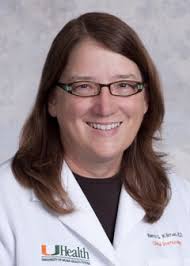
Moderators: Nancy Klimas, M.D., FACP, FIDSA, Chair, Department of Clinical Immunology, Director, Institute for Neuro-Immune Medicine, Nova Southeastern University and Theresa Michele, M.D., Clinical Team Leader, Division of Pulmonary, Allergy, and Rheumatology Products (DPARP), Office of Drug Evaluation II (ODE II), Office of New Drugs, CDER, FDA

Panelists: Lucinda Bateman, M.D., Fatigue Consultation Clinic, Salt Lake City, Utah, Lisa W. Corbin, M.D., FACP, Associate Professor, Division of General Internal Medicine University of Colorado Denver School of Medicine, Lily Chu, M.D., MSPH, International Association for CFS/ME and Patient, Jose G. Montoya, M.D., FACP, FIDSA, Professor of Medicine, Division of Infectious Diseases and Geographic Medicine Stanford University School of Medicine. Jennifer Spotila, JD, Patient and Christine Williams, MEd, Patient.

Questions: What were your key take-away messages from the discussion yesterday on the most significant symptoms experienced by patients with CFS and ME? Please describe any significant differences in your experience as clinicians and patients compared to yesterday’s discussion. [timestamp 8:44 – 24:00]

Dr. Bateman said that she concurred with what she heard yesterday. She would add the psychological suffering that comes along with the illness. Top of the list are cognitive limitations and fear.

Dr. Lily Chu talked about the results of the survey she conducted. Fatigue, PEM (post-exertional malaise), pain, and cognitive impairments came first. Multiple chemical sensitivities, gastro-intestinal symptoms, and orthostatic intolerance appeared in over 50% of respondents. The impact of the illness was substantial: respondents were more than 95% impaired and 89% needed assistance for daily chores.

Dr. Montoya stated that the two most salient clinical features were cognitive dysfunction and unpredictable crashes. The ability to drive is significantly impaired in his patients. This makes sense, because driving is the perfect example of multi-tasking. When patients get better they start to come to their visits alone. Herpes simplex is a trigger in a cohort of patients. The third thing to consider is that there are several triggers for patients. We should keep in mind how the effect of a drug is undermined when there are several triggers.

Christine Williams, who became ill at the age of 56, brought up the “double whammy.” These are the effects of this illness on an aging population. For her, the flu-like symptoms are the worst, because they make her feel very sick.

Jennie Spotila: We need to remember that the working ill, who struggle to maintain jobs and raise families, are a significant part of the patient community. For her, the words “fatigue” and “tired” are placeholders to avoid the long description of a crash. She stressed the need to include multiple aspects of cognitive function in order to measure improvement in drug trials. Ms. Spotila noted that gut disturbances had not been addressed. She also pointed out that prevalent symptoms, like orthostatic intolerance, can be masked. In addition, many symptoms could be measured and identified, but people are not looking at them. There is a lot of suffering that is going under the radar.

Question 2: Based on your expertise as clinicians and your experience as patients, which symptoms of CFS and ME could be identified as valid, quantifiable and reliable outcome measures or endpoints in clinical trials to evaluate potential drugs to treat CFS and ME? [timestamp 24:01 - 39:10]

Dr. Lily Chu: Any symptom of ME could be used as a valid and quantifiable outcome measure, it depends on which scales you want to use and which drug you want to test. She also mentioned that because the disease is heterogeneous, studying subgroups would yield better results. Her second point is that it is very important to come up with a good outcome measure for post-exertional malaise. Post-exertional malaise (PEM) is not that same as post-exertional fatigue. Malaise can have a flu-like component, and involves collapse and confusion. There is not enough basic research on PEM. The difference between fatigue and fatigability is important, because it not only takes into account the subjective feeling of feeling tired, but puts it in relation to activities. The real test is whether a drug improves function, rather than measuring a subjective state.

Dr. Montoya
Dr. Montoya talked about the importance of separating out symptoms. He mentioned a double-blind study he did in whicha smallgroup of patients were given antivirals. Their cognitive symptoms improved before their physical symptoms, but this improvement was only discovered when they compared a subscore on their initial questionnaire. Otherwise, it may have been missed. Separating cognitive fatigue from physical fatigue could be helpful for clinical trials. Because of the fluctuating nature of the disease he recommended using linear regression statistical models in order to measure various points, rather than simply comparing a beginning and endpoint. Type of onset is also extremely important. Do patients have viral onset, abrupt onset? What is the duration of the illness? “It’s very hard to believe that a patient with CFS of two years’ duration would have the same pathogenesis as a patient with 20 years’ duration”.

Dr. Lisa Corbin brought up the point that while it is important to look at function as an endpoint, there may be different parameters for function in the CFS/ME population. For example, being able to read a book for two hours (or at all) will not appear on any functional scale. The way we measure function needs to be more specific for this population. It’s important to follow the symptoms that are most distressing for patients.

Dr. Bateman mentioned that there are already objective measures in place to measure orthostatic intolerance,
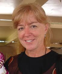
Dr. Bateman
sleep disorder, as well as other symptoms. One problem in clinical testing is that as a patient’s ability to function increases they do more, which produces symptoms. So, unless patients maintain the same level of activity throughout the trial, measuring symptoms is unreliable.

III. Question 3: What are the key factors you take into account when making decisions to prescribe (as clinicians) or use (as patients) therapies to treat symptoms associated with CFS and ME? [timestamp 40:00 - 56:15]

Dr. Lisa Corbin: The first factor is patient input. I treat the most disabling symptom first. I do treatments one at a time. Start low and go slow.

Dr. Bateman talked about individualizing treatments as well as managing and coping strategies.

Dr. Montoya talked about preventing the wild fluctuations of the illness. He also addressed treating infectious triggers. He does PCR tests for HHV6 and measures viral loads. Those patients are treated separately. Other patients undergo testing to identify other viral triggers, risk factors for Q fever, and other pathogens. But, Dr. Montoya stressed, the pathogens must be correctly identified.

Question 4: If you had a drug to explore what would be your top one or two choices? [timestamp 56:26 – 58:56]

Dr. Bateman: On top would be antiviral and immune-modulating drugs. Next would be symptom management drugs for pain, stimulants for cognition, and sleep meds and meds for OI (orthostatic intolerance).

Dr. Lily Chu: Same plus Rifaxamin

Dr. Montoya: Antivirals, immune modulators. He believes that the immune response to the infectious agent is what drives CFS/ME symptoms, but for some patients it may be too late.

Jennie Spotila added Rituximab, as well as drugs that can increase VO2 max. These could potentially make a huge difference in the functionality of patients.

Question and Answer Period [timestamp 58: 56]

Question: How do we deal with the fact that so many patients are not located close to clinical trials?

Answer: Dr. Klimas mentioned that there is a spouse CBT protocol that involves long-distance CBT via a video group session. (Dr. Klimas noted that CBT is not a treatment.) Dr. Klimas added that there is a disconnect between 20-year-old guidelines and how doctors actually treat their patients.

[Questions about viral makers and immune markers were referred to, but the questions were not read.]

Question for Dr. Montoya:  “Do you subgroup out to foreign diseases, and what is the prevalence of HSV and CMV in your practice, and do you use cidofovir (Vistide) for HSV positive patients?

Answer: Dr. Montoya: We have a small group of patients with positive test results, but it is hard to come up with treatments for tick-borne illnesses. For herpes simplex we use acyclovir. We reserve cidofovir for patients who are severely ill, because it is very toxic. Doctors need to remember to “do no harm.”

Question: How can we better break down the barriers of data sharing?

Answer: Lily Chu: There are problems with privacy when it comes to freely disseminating medical data from individual patients. Dr. Montoya: Something similar needs to be done as was done for AIDS. Within 15 years they had highly effective treatments. We need a “bailout” for CFS/ME, a huge infusion of money and infrastructure. Dr. Vernon mentioned that CAA requires that anyone they give a grant to must share their data. Dr. Klimas stated that NOVA shares its data with other CFS/ME institutions, and there are other multi-institutional initiatives underway. Dr. Montoya mentioned that Stanford also has a biobank of several hundred CFS/ME patients. There is, in fact, a large amount of data already gathered and accessible for pharmaceutical companies.

Originally posted on ProHealth.
Related Posts Plugin for WordPress, Blogger...